
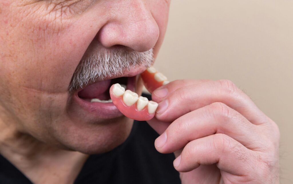
Denture sores are one of the most common complaints among denture wearers. Whether you have just received new dentures or have worn them for years, painful spots on the gums, roof of the mouth, or the tissue under your dentures can develop from a range of causes. Understanding what denture sores look like, what causes them, and how to treat them quickly makes a significant difference in both comfort and long-term oral health. This guide covers the full picture — from the clinical appearance of different types of denture sores and denture stomatitis to home treatment, the fastest ways to heal, and when professional dental care is necessary.
What Do Denture Sores Look Like?
Denture sores vary considerably in appearance depending on their cause, severity, and location. The following descriptions cover the main types you may notice in the mirror or when examining your mouth.
Pressure Sores
Pressure sores are the most common type of denture sore and appear as red, tender, well-defined spots on the alveolar ridge — the bony arch of gum tissue where the denture sits. The redness is concentrated at the exact point where the denture edge or base presses most firmly against the tissue. In early stages they appear as bright red, slightly raised spots. If the pressure continues without relief, the centre of the sore may develop a small shallow ulcer with a pale yellow or whitish base surrounded by red inflamed tissue.
Pressure sores are most commonly found along the ridge of the lower jaw where lower dentures sit, and along the hard palate and upper ridge where upper dentures contact the tissue.
Traumatic Ulcers
Traumatic ulcers from denture friction are open sores — usually round or oval — with a shallow crater, a pale or yellowish floor, and a clearly defined red inflamed border. They are typically 2 to 10 millimeters in diameter. They bleed easily when touched or when eating, and the surrounding tissue is tender and swollen.
Unlike canker sores (aphthous ulcers), which appear spontaneously and resolve on their own, denture-related traumatic ulcers persist as long as the denture continues to create friction in the same location. Removing the denture and allowing the tissue to rest is the first step in allowing them to heal.
Denture Stomatitis
Denture stomatitis is a specific condition — also called prosthetic stomatitis or Candida-associated stomatitis — that affects the tissue covered by the denture, most often the palate (roof of the mouth) under an upper denture. It is caused primarily by Candida albicans, a fungal organism that colonizes the fitting surface of dentures and the underlying oral mucosa.
Denture stomatitis appears as diffuse redness of the palatal mucosa — the tissue appears uniformly red, inflamed, and sometimes mildly swollen, covering the entire area in contact with the denture rather than appearing as isolated spots. There are three clinical types classified by severity. Type I (pinpoint hyperemia) appears as small, scattered red spots. Type II (diffuse erythema) covers the full denture-bearing area with uniform redness. Type III (granular or inflammatory papillary hyperplasia) presents as a bumpy, cobblestone-textured red surface in chronic cases.
Denture stomatitis is often painless in its early and moderate forms, which means many wearers do not seek treatment until the condition is well established. It affects an estimated 65 percent of denture wearers to some degree, making it the most prevalent oral condition associated with denture use.
Allergic Reaction Sores
Allergic reactions to denture materials — most commonly to residual monomer in acrylic resin, nickel in metal components, or specific denture adhesive ingredients — produce a distinct pattern of redness and swelling that covers the entire area contacting the allergenic material rather than pressure points specifically. The tissue appears uniformly red and may be swollen, with possible small vesicles (fluid-filled bumps). The condition often produces a burning sensation or itching rather than the sharp pain of pressure sores.
True allergy to dental acrylic is relatively uncommon — most reactions to denture materials are contact irritation rather than true allergic (IgE-mediated) reactions. A dentist can distinguish between the two through the pattern of tissue involvement and history.
Angular Cheilitis
Angular cheilitis — cracks, redness, and sometimes crusting at the corners of the mouth — is associated with denture wear, particularly when dentures have lost vertical height (the teeth have worn down, causing the mouth to close too far). The reduced vertical dimension creates deep folds at the mouth corners that collect saliva and provide a warm, moist environment for Candida and bacterial colonization.
Angular cheilitis appears as red, raw, sometimes cracked or encrusted fissures at one or both corners of the mouth. It is often associated with denture stomatitis and can be treated with antifungal cream alongside denture adjustment to restore correct vertical height.
Denture Stomatitis Pictures: What to Look For
When self-examining for denture stomatitis, look for the following visual indicators in a well-lit mirror after removing your dentures.
- Uniform pink-to-red discoloration of the palate covering the area where the denture base sits — distinct from the normal pale pink color of healthy oral mucosa
- The redness is confined to the denture-bearing area with a relatively sharp border where the denture edge rests
- In more advanced cases, the tissue may have a bumpy, granular, or cobblestone texture — this is inflammatory papillary hyperplasia (Type III denture stomatitis)
- White patches on the palate — particularly those that wipe off leaving a red raw surface — may indicate candidiasis (thrush) associated with denture stomatitis
- The fitting surface of the denture may have a whitish or yellowish biofilm coating if not cleaned thoroughly
Important: White patches in the mouth that cannot be wiped off and persist for more than two weeks should always be evaluated by a dentist or physician. White patches (leukoplakia) can occasionally be a sign of pre-cancerous changes and require professional assessment.
Causes of Denture Sores
Ill-Fitting Dentures
The most common cause of denture sores is dentures that no longer fit correctly. Denture fit deteriorates over time because the alveolar bone and gum tissue beneath dentures gradually resorbs — this is a natural biological process that occurs when teeth are absent, as the bone no longer receives the stimulation it needs to maintain its volume. The rate of resorption is highest in the first year after tooth extraction and continues, more slowly, throughout life.
As the bone resorbs, dentures that once fit precisely begin to sit differently — rocking on the ridge, concentrating pressure on specific points, and creating friction in areas that were previously well-cushioned. The ADA recommends denture evaluation every one to two years to assess fit changes.
Poor Denture Hygiene
Inadequate denture cleaning allows the accumulation of Candida albicans on the fitting surface of the denture — the primary cause of denture stomatitis. The porous acrylic material of denture bases harbors microorganisms in microscopic surface irregularities that are not removed by rinsing alone. Biofilm (plaque) on the denture surface continuously exposes the underlying tissue to fungal and bacterial irritants.
Dentures should be cleaned daily with a soft brush and non-abrasive denture cleaner or mild dish soap — never regular toothpaste, which is too abrasive for acrylic and scratches the surface, creating more sites for microbial colonization.
Wearing Dentures Around the Clock
Wearing dentures continuously — including overnight — significantly increases the risk of denture stomatitis and pressure-related sores. Removing dentures for at least six to eight hours daily (typically overnight) gives the underlying tissue a period of recovery and reduces the total time Candida is in contact with the palatal mucosa. Studies consistently show that continuous denture wear is the strongest predictor of denture stomatitis severity.
New Dentures
New dentures routinely cause some degree of pressure sores during the adjustment period — even accurately made dentures require a break-in period as the tissue adapts to the new pressure distribution. Most dentists advise new denture wearers to expect some soreness in the first two to four weeks and schedule adjustment appointments to relieve pressure points identified by the pattern of sores.
Dry Mouth
Saliva plays a critical role in protecting oral tissues from denture friction — it acts as a lubricant between the denture surface and the mucosa. Dry mouth (xerostomia), often a side effect of medications including antihistamines, antidepressants, antihypertensives, and diuretics, significantly increases friction and sore formation. More than 400 medications list dry mouth as a side effect.
Bone and Tissue Changes
Beyond the gradual resorption that affects all denture wearers, specific changes can accelerate fit problems. Weight loss reduces the soft tissue padding under dentures. Certain medications (particularly long-term corticosteroids) can affect bone density. Osteoporosis is associated with accelerated alveolar bone loss. Medical conditions including diabetes increase susceptibility to oral infections and impair tissue healing, making denture sores more likely and slower to resolve.
Types of Denture Sores at a Glance
| Type | Appearance | Location | Primary Cause | Pain Level |
| Pressure sore | Red spot, may develop shallow ulcer | Ridge, denture edge contact points | Ill-fitting denture | Moderate to high |
| Traumatic ulcer | Open sore, pale base, red border | Any friction point | Denture rubbing | High, bleeds easily |
| Denture stomatitis | Diffuse redness, may be bumpy | Palate (upper), full bearing area | Candida + poor hygiene | Low to none (often painless) |
| Allergic reaction | Uniform redness, swelling, possible vesicles | Full contact area | Material sensitivity | Burning/itching |
| Angular cheilitis | Red cracks at mouth corners | Lip corners | Candida, reduced vertical height | Low to moderate |
Fastest Ways to Heal Denture Sores
The fastest healing path for any denture sore is removing the source of irritation first, then supporting tissue recovery.
Remove the Denture
Remove the denture completely for several hours each day, and overnight without exception. This is the single most effective step for allowing sore tissue to begin healing. Pressure sores and traumatic ulcers cannot heal while the denture continues to press on them.
Warm Salt Water Rinses
Dissolve half a teaspoon of salt in eight ounces of warm water and rinse for 30 to 60 seconds, three to four times daily. Salt water is mildly antiseptic, reduces bacterial load around the sore, reduces swelling through osmotic effects, and creates an environment that promotes tissue healing. It is safe for daily use and is the most recommended home care for oral sores.
Topical Oral Analgesics
Over-the-counter oral analgesics containing benzocaine (such as Orajel or Anbesol) can be applied directly to painful sores to provide temporary pain relief before eating or wearing the denture. These products numb the surface tissue temporarily. They do not speed healing — they manage pain while the tissue recovers.
Aloe Vera Gel
Pure aloe vera gel applied to oral sores has some evidence supporting its anti-inflammatory and tissue-soothing properties. A study published in the Journal of Dentistry found aloe vera effective in reducing pain and promoting healing of traumatic oral ulcers. Use only food-grade or medical-grade aloe vera gel in the mouth, not products containing additives, colorings, or alcohol.
Antifungal Treatment for Denture Stomatitis
Denture stomatitis caused by Candida does not resolve with salt water rinses or rest alone — it requires antifungal treatment. Nystatin oral suspension (swish and spit) or clotrimazole troches (lozenges) are the standard first-line treatments and are available by prescription. Miconazole oral gel is available over the counter in some formulations.
Crucially, the denture itself must also be treated — Candida in the acrylic biofilm will reinfect the tissue if the denture is not disinfected. Soaking dentures in a dilute chlorhexidine solution or a commercial denture disinfectant overnight alongside antifungal treatment is necessary for effective resolution.
Denture Adjustment
For pressure sores and traumatic ulcers caused by ill-fitting dentures, a dentist can perform a denture adjustment — identifying the pressure point causing the sore using disclosing paste and selectively relieving the acrylic in that area. This is the only way to address the underlying mechanical cause. A well-adjusted denture allows sores to heal fully and prevents recurrence.
When to See a Dentist for Denture Sores
Home care measures can relieve symptoms and support healing, but professional attention is necessary in the following situations.
- The sore has not improved after one to two weeks despite removing the denture for rest periods and using home care measures
- The sore is worsening rather than improving
- There are signs of infection: increasing redness, warmth, swelling, pus, or fever
- The sore makes eating, drinking, or speaking impossible
- You have diffuse palatal redness suggesting denture stomatitis — antifungal treatment is required
- Any white patch in the mouth that does not wipe off and has been present for more than two weeks — this requires professional evaluation to rule out pre-malignant changes
- You have diabetes, are immunocompromised, or take medications that reduce immunity — oral infections can escalate more quickly in these patients
How to Prevent Denture Sores
Daily Denture Cleaning
Clean dentures thoroughly every day using a soft denture brush and a non-abrasive denture cleanser or mild liquid soap. Brush all surfaces including the fitting surface that contacts the gum tissue. Rinse thoroughly after cleaning. Soak dentures overnight in a denture cleaning solution to disinfect and remove biofilm that brushing alone cannot eliminate.
Never use regular toothpaste on dentures — its abrasive particles scratch the acrylic surface, creating microscopic grooves that harbor bacteria and Candida and accelerate stomatitis. Never use bleach on metal-containing dentures.
Remove Dentures at Night
Removing dentures for at least six to eight hours every day — ideally overnight — is the most important single habit for preventing denture stomatitis. This rest period allows the palatal mucosa to recover from the constant pressure and microbial exposure of daytime wear and significantly reduces Candida colonization. Store dentures in clean water or denture solution while they are out.
Regular Dental Check-Ups
The ADA recommends that denture wearers see their dentist at least once a year, and every one to two years for formal denture evaluation. Regular appointments allow early identification of fit changes before they cause sores, professional cleaning of the denture, and assessment of the oral tissue for any concerning changes. Many people avoid dental visits after receiving dentures, which is why denture stomatitis often goes undiagnosed and untreated for years.
Address Dry Mouth
If you take medications that cause dry mouth, discuss the issue with your prescribing physician — alternative medications with less xerostomic effect may be available. Staying well hydrated, using xylitol-containing products, and using over-the-counter saliva substitutes or mouth moisturizing gels can reduce the friction that causes sores in dry mouth patients.
Consider Denture Relining or Replacement
Dentures that cause recurring sores despite adjustments may need relining — the addition of new material to the fitting surface to restore accurate contact with the changed tissue — or full replacement. Complete dentures typically last five to seven years before the changes in the underlying bone and tissue make them sufficiently ill-fitting to warrant replacement. Wearing worn-out dentures to avoid the cost of replacement is one of the most common causes of chronic denture sores and bone loss acceleration.
Frequently Asked Questions
What do denture sores look like?
Denture sores vary by type. Pressure sores appear as red, tender spots on the gum ridge at contact points. Traumatic ulcers are open sores with a pale or yellowish floor and a red border. Denture stomatitis appears as diffuse redness covering the palate under an upper denture — often uniform and covering the full denture-bearing area. Allergic reactions produce widespread redness and possible swelling. Angular cheilitis appears as cracks and redness at the mouth corners.
How long do denture sores take to heal?
Minor pressure sores typically improve within three to seven days once the source of pressure is removed through rest or denture adjustment. Larger traumatic ulcers may take seven to fourteen days to heal fully. Denture stomatitis (Candida-associated) requires antifungal treatment and improved hygiene to resolve — without treatment it does not heal spontaneously. Any sore that has not improved after two weeks requires professional evaluation.
What is the fastest way to heal denture sores?
The fastest healing path is: remove the denture for rest, rinse with warm salt water three to four times daily, and apply topical analgesic for pain relief. For pressure sores caused by fit problems, a dentist’s adjustment removes the pressure point and allows the tissue to heal fully rather than continuing to irritate it. For denture stomatitis, antifungal medication alongside denture disinfection resolves the condition; salt water alone is insufficient.
Can you get sores from new dentures?
Yes — pressure sores from new dentures are entirely normal and expected, particularly in the first two to four weeks of wear. Even accurately made dentures create some pressure points that require adjustment. Most dentists include adjustment appointments in the cost of new dentures precisely for this reason. Report sore spots to your dentist at your post-fitting appointments so pressure points can be identified and relieved.
How do I know if my denture needs adjusting?
Signs that your denture needs adjustment include recurring sores in the same location, dentures that rock or shift during eating or speaking, difficulty chewing on both sides evenly, clicking sounds when closing, and dentures that feel loose even with adhesive. Changes in the shape of your face — particularly increased vertical skin folds around the mouth — can indicate loss of vertical dimension from worn teeth and may require new dentures rather than simple adjustment.
Should I sleep with my dentures in?
No — denture wearers should remove dentures for sleep. Continuous wear significantly increases the risk of denture stomatitis and pressure sores. The ADA and most dental authorities recommend removing dentures for at least six to eight hours every day. Removing dentures overnight also allows the jawbone and gum tissue a period of pressure relief that supports long-term tissue and bone health. Store dentures in water or denture solution when not being worn.
Final Thoughts
Denture sores are common but not inevitable — and they are not something you simply have to tolerate. Understanding what different types of denture sores look like, identifying their cause, and taking the appropriate steps to remove the source of irritation resolves most cases. Persistent sores, recurring sores in the same location, or any oral lesion that does not heal within two weeks should always be professionally evaluated.
This article is for informational purposes only. If you have denture sores that are not improving or that concern you, consult a licensed dentist for an examination and personalized treatment recommendations.
DISCLAIMER: This article is for informational purposes only and does not constitute professional dental or medical advice. If you have persistent denture sores or oral lesions, consult a licensed dentist for diagnosis and treatment.